


X-ray diagnostics
X-rays remain important today for assessing the fine structure of bones, complementing other diagnostic methods. In specific cases, such as determining whether there is a calcium deposit in the shoulder joint, an X-ray is a useful addition to MRI.
Of course, radiation exposure can be minimized through various measures and is usually relatively low in these recordings.
Calcium deposit in a shoulder joint

This X-ray of a right shoulder joint shows a large oval calcium deposit approximately in the center of the image. X-rays are sometimes better suited than MRI scans for detecting such calcifications.
Cervical spine - overview image
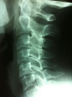
This lateral x-ray image of a cervical spine shows a degenerative change in the intervertebral disc between the 5th and 6th cervical vertebrae.
Online appointment
- Schedule now
Im Zollhafen 6, 50678 Köln, Deutschland
