


Meniscus repair - Meniscus tear treatment without surgery
The meniscus in the knee joint is an important structure, functioning as a shock absorber and guiding the movement of the cartilage surfaces. Therefore, every effort should be made to preserve the meniscus if it is injured.
Watch a video about our new treatment method
In our practice, we have developed a modern, minimally invasive treatment concept that aims not to remove a meniscus tear, but to preserve it and bring it to healing.
The focus is on the gentle refixation of the meniscus tear, allowing the tissue to heal. The procedure is performed on an outpatient basis, without surgery or general anesthesia – instead, it is carried out under local anesthesia and precise MRI guidance. This makes the treatment virtually painless and, at the same time, extremely accurate. The procedure takes approximately 30 to 60 minutes.
A major advantage for our patients: The knee can usually be put under stress again immediately afterwards, thus avoiding longer periods of downtime.
To support the healing process, a biological building phase with the body's own growth factors follows. These are obtained from a single blood draw and then injected once a week for four weeks, specifically near the meniscus tear – also under local anesthesia.
After about six weeks, the healing process is usually complete and is finally checked using an MRI scan.
Our goal is to preserve the meniscus, maintain the natural function of the knee joint, and enable the quickest possible return to everyday life and sporting activities.
Using this procedure, we have successfully performed over 1090 treatments since mid-2016, and the therapeutic results have been very promising. We presented the meniscus repair method at the international EFORT Congress in Vienna (May 31, 2017). The first treatment results are now available one year after meniscus refixation. All patients examined after one year were doing well and were largely symptom-free, except for five patients who subsequently underwent surgery for the meniscus. We presented the results one year after refixation at the international meeting of the ICRS (International Cartilage Repair Society) in Milan in December 2018. The follow-up results after four years are now available and were published in the journal Sports Injuries & Medicine, Volume 5, Issue 1. These results demonstrated a significant improvement in all WOMAC scores even four years after treatment.
The treatment results have now also been published in a new book (03.2023):
Research Highlights in Disease and Health Research Vol. 5
Buch-Kapitel Nr. 4: Improved Treatment of Knee Meniscus Injuries and Reduced Need for Surgery with a Combination of Tisseel Fibrin Glue and Orthokine ACS: A 4-Year Follow-Up Archive Study
Results of our treatment method after 4 years
View the results here
The following illustrations show the principle of the minimally invasive treatment method:
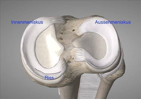
Fig. 1) Shows a top view of a right knee joint with the medial and lateral menisci. A typical tear is shown in the medial meniscus (bottom left in the image).
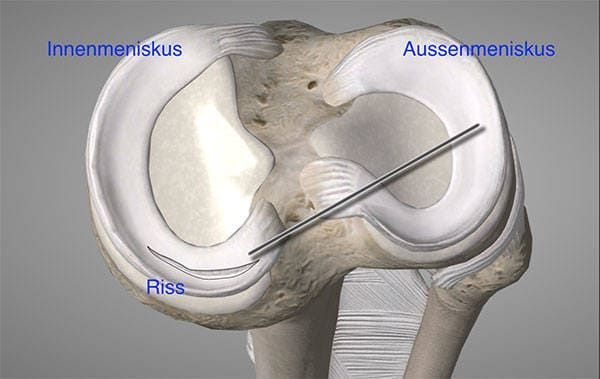
Fig. 2) Shows the insertion of an MRI-compatible introducer under MRI guidance into the meniscus tear.
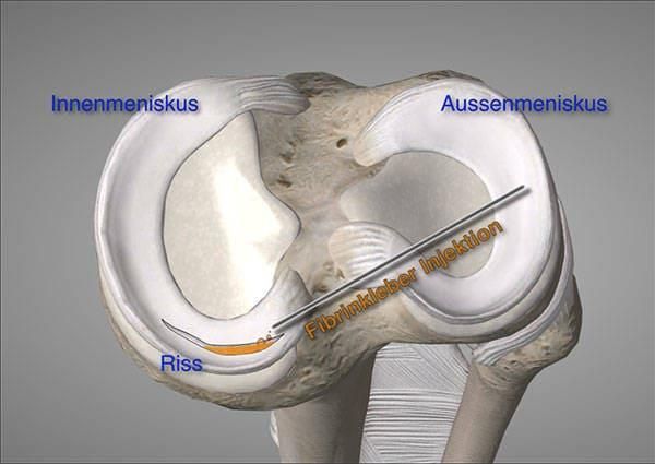
Fig. 3) After checking the position of the introducer in the MRI, the fibrin glue is injected.
Advantages of the new method:
1. No surgical risk: Since no surgery is required, the associated surgical risk is completely eliminated. Even minimally invasive procedures such as arthroscopic surgery carry the risk of cartilage damage as well as injuries to nerves or blood vessels – these are avoided here.
02. No anesthesia: General or spinal anesthesia is not necessary, thus completely eliminating the risks of anesthesia.
03. Preservation of the meniscus: The meniscus remains fully intact as a central structural element in the knee joint – an important contribution to long-term joint stability and function.
4. Virtually painless treatment: The method is generally associated with very little pain for patients.
5. No radiation exposure: The procedure is performed under MRI (magnetic resonance imaging) guidance, thus avoiding exposure to X-rays.
6. Immediate load-bearing capacity: The muscles are not weakened, so direct weight-bearing on the knee is possible. Furthermore, the time commitment for the patient is comparatively short.
7. Long-established material: The fibrin glue used is a proven product that has been successfully used in medical practice for over 29 years.
Watch a video about our new treatment method
Results of our treatment method after 4 years
View the results here
The treatment results have now also been published in a new book (03.2023):
Research Highlights in Disease and Health Research Vol. 5
Buch-Kapitel Nr. 4: Improved Treatment of Knee Meniscus Injuries and Reduced Need for Surgery with a Combination of Tisseel Fibrin Glue and Orthokine ACS: A 4-Year Follow-Up Archive Study
FAQs on the refixation of tissue fragments with fibrin glue (Tisseel) and autologous conditioned serum (ACS)
Is the procedure limited to certain age groups?
- No, the procedure is applicable regardless of age.
What can you glue with Tisseel? *
- Shoulder joint: Shoulder tendons (supraspinatus tendon tears, subscapularis tendon tears, labral tears)
- Foot: Achilles tendon ruptures, plantar fascia ruptures
- Wrist: Scapholunate band
- Elbow joint: Collateral ligament
- Knee joint: Meniscus, patellar tendon, quadriceps tendon
* The assessment will be carried out taking into account the crack formation and the respective circumstances, using Mr. Strümper's current MRI findings as a basis.
Can it also be used to glue cartilage defects?
- Pro-inflammatory cytokines released from damaged cartilage areas lead to inflammatory reactions and associated pain in the surrounding tissue. - To stabilize and seal the cartilage defects, fibrin glue (e.g., Tisseel) can be used. Subsequently, autologous conditioned serum (ACS) is applied to promote structural regeneration through growth-stimulating factors.
Therefore, we use targeted injections of autologous conditioned serum (ACS) because spontaneous healing is not possible: Highly differentiated cartilage cells are unable to produce growth factors themselves. Furthermore, there are no blood vessels in cartilage tissue that could transport growth factor-producing cells, such as monocytes, to the defect area.
How long does the treatment take in total?
The treatment lasts approximately 6 weeks; during this time, you simply need to refrain from sporting activities. Afterwards, muscle-strengthening exercises as part of physiotherapy are recommended.
Is it necessary to preserve the meniscus?
- The meniscus in the knee joint is an important organ and functions as a kind of shock absorber and guides the movement of the cartilage joint surfaces.
Therefore, every effort should be made to preserve the meniscus if it is injured.
Will the procedure take place under general anesthesia?
- No, the procedure takes place under local anesthesia and MRI guidance.
What happens after the 6 weeks?
- Following refixation and infiltration therapy with ACS, medical physical treatment and appropriate muscle strengthening training are recommended.
How is the procedure carried out?
- Tissue bonding is performed without surgery under local anesthesia and MRI guidance using a fibrin adhesive (Tisseel), followed by the promotion of healing through four to six weekly injections of growth factors (ACS).
What does the entire 6-week treatment include?
- Preliminary consultation/explanation, clinical examination
- ECG measurement
- Blood collection for the production of autologous conditioned serum (ACS)
- Refixation with Tisseel in open MRI (approx. 1 hour) followed by a stay in the recovery room (approx. 30 minutes)
- Patient check-up/discharge (if necessary, loan of crutches for stabilization)
- The next day or follow-up MRI the day after (if necessary, return crutches 1 day after refixation)
- 1 week later or 4-6 ACS injections at weekly intervals (earliest start: 1 week after refixation)
- Completion of therapy or follow-up MRI 6 weeks after refixation / final consultation
Why is the ACS necessary for the procedure?
- Infiltration therapy with ACS following refixation is necessary because the adhesive slowly dissolves after 6 weeks. To stimulate and support the timely fusion of the tissues, we use the biological factors (various cytokines, especially growth factors) present in high concentrations in ACS, which have a regenerative effect on the tissue.
How does the procedure with the ACS work?
- For ACS therapy, 20ml of blood is drawn from the patient in a single procedure. A serum enriched with growth factors is extracted from this blood using a special process, which we then apply directly to the defect deep within the joint under MRI guidance for precise infiltration.
What is ACS?
ACS/BCS is produced from venous blood collected using an EOT®II syringe (Orthogen Lab Services, Germany). The patient's blood is incubated in the device for 6 hours at 37°C, and the serum is then obtained by centrifugation. This serum is injected into the patient's joint at weekly intervals. The conditioned, cell-free serum is rich in the growth factors IGF, TGF-β, EGF, FGF, PDGF, and the cytokine IL-1Ra. It also contains smaller amounts of IL-1β, IL-6, and TNF-α [33].
Is there anything I need to be aware of when having my blood drawn for the ACS test?
- When taking blood to obtain autologous conditioned serum (ACS), you should pay attention to the following:
To obtain enough serum of the best quality, you should avoid caffeine, alcohol and theine for 3 days before the blood draw and drink plenty of water instead.
Once the blood sample has been taken, the preparations are complete and you can resume your usual routine. On the day of the blood draw, you can eat breakfast as normal and do not need to arrive on an empty stomach.
What risks might I encounter?
As with any procedure, there are certain risks which we naturally keep as low as possible, such as:
- Risk of infection
- Allergy, or allergic reaction
- New treatment method, long-term results not yet available
Why is the procedure performed under MRI?
- Visual control during precise refixation and infiltration therapy in and around a lesion deep within the joint is best achieved using magnetic resonance imaging (MRI).
MRI offers a particularly excellent opportunity due to its three-dimensional capabilities.
Topographic live representation, the representation of the injection needle within the anatomical structures, especially arteries and nerves.
Another advantage is that, unlike X-ray or CT scans, MRI is unrestricted and completely radiation-free. Ultrasound makes it difficult to accurately visualize the structures deep within the joint and cannot be performed under the same sterile conditions.
Will I need to use forearm crutches after the refixation?
The adhesive fixes the torn meniscus so effectively that full weight-bearing is possible immediately after the procedure. Therefore, the risk of thrombosis with this method is minimal. We recommend using forearm crutches on the day of the refixation.
These are only for support, as your leg may become temporarily numb due to the local anesthetic. You will no longer need the forearm crutches from the following day.
Can I drive again on the day of the meniscus refixation?
- On the day of your meniscus repair, it is best to bring someone with you who can pick you up after the procedure, as your foot may become numb due to the local anesthetic, making you unable to drive.
How can the meniscus heal in this situation?
Fibrin glue is able to fix and adapt the meniscal tear. Similar to arthroscopic suturing or pin fixation, this aims to facilitate cellular healing. In a damaged joint, the environment is often inflammatory/catabolic. Augmentation with the growth factors ACS/BCS has proven effective in a number of experimental and clinical studies with degenerative and acute meniscal tears.
Tissue injuries have been shown to have anti-inflammatory and healing properties. It should be able to support healing more efficiently if the edges of a meniscal tear are kept in close contact long enough to allow cell contact/healing and fusion. This procedure has been performed in an environment where anesthesia and surgery could be avoided. It may be suitable for a subset of injuries where surgical intervention is not strictly necessary.
How successful is the treatment?
- According to our most recent study (as of February 2025), after treatment of over 990 people, approximately 82% of the respondents are symptom-free or have only very minor symptoms.
! Often, pre-existing conditions (osteoarthritis, joint cartilage damage, etc.) can lead to persistent symptoms that are not attributable to the glued meniscus!
Is it possible to shorten the procedure?
Unfortunately, it is not possible to shorten the procedure, as weekly injections of growth factors (ACS) after gluing are crucial for the healing process. Shortening the procedure could lead to the refixed area not healing properly, which would jeopardize the success of the procedure.
- It is not possible to split the treatment, for example, to perform the refixation in our practice and the ACS injections externally.
Is it possible to interrupt the proceedings?
- An interruption of the procedure is generally not planned. It is important that the first injection takes place exactly one week after the bonding.
- Should an interruption occur later for any reason, this is possible for a maximum of one week – longer breaks should be avoided.
What is fibrin glue made of?
The fibrin sealant is obtained from the blood plasma of selected donors. Similar to the transfusion of donor blood, numerous tests are mandatory. These ensure that the donor plasma is not contaminated with bacteria, hepatitis, or AIDS viruses.
- Fibrin glue is a biological tissue adhesive consisting of two components: one
Fibrinogen solution (containing fibrinogen, factor XIII, and apoprotein) and a thrombin solution (containing thrombin and calcium chloride). When mixed, fibrinogen is converted into fibrin by thrombin, forming a stable adhesive that stops bleeding and binds tissues together.
The apoprotein inhibits fibrin-degrading enzymes and thus stabilizes the adhesive. Its components are usually derived from human plasma or produced recombinantly.
How much would the procedure cost?
- The total cost for the entire treatment scope of a classic refixation with Tisseel and subsequent ACS therapy is approximately €4,500 - €5,000 (material costs may vary).
- In the event of additional necessary measures, additional costs may be incurred.
Will the costs of the procedure be covered by the private insurance?
- Please note that not every private insurance company covers the costs of the procedure.
Therefore, we will provide you with a cost estimate that you can submit to your insurance company. This will make it easier for you to clarify the approval for coverage of the costs of the procedure.
Patient testimonial after meniscus treatment with fibrin glue (video)
Results of our treatment method after 4 years
View the results here
Comparison images of a torn medial meniscus of a right knee joint over time before and after refixation with fibrin glue in MRI.
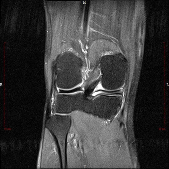
Before the procedure
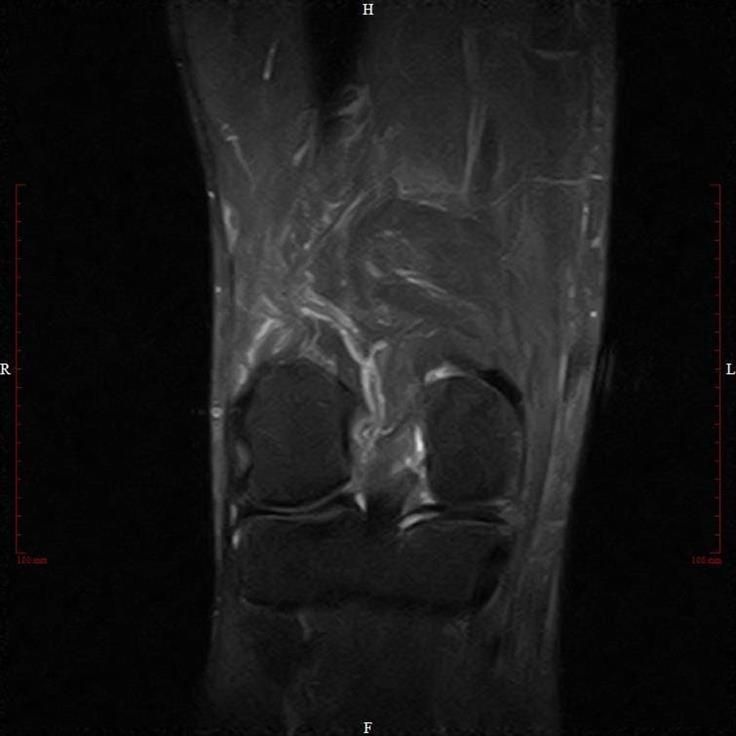
6 weeks after refixation
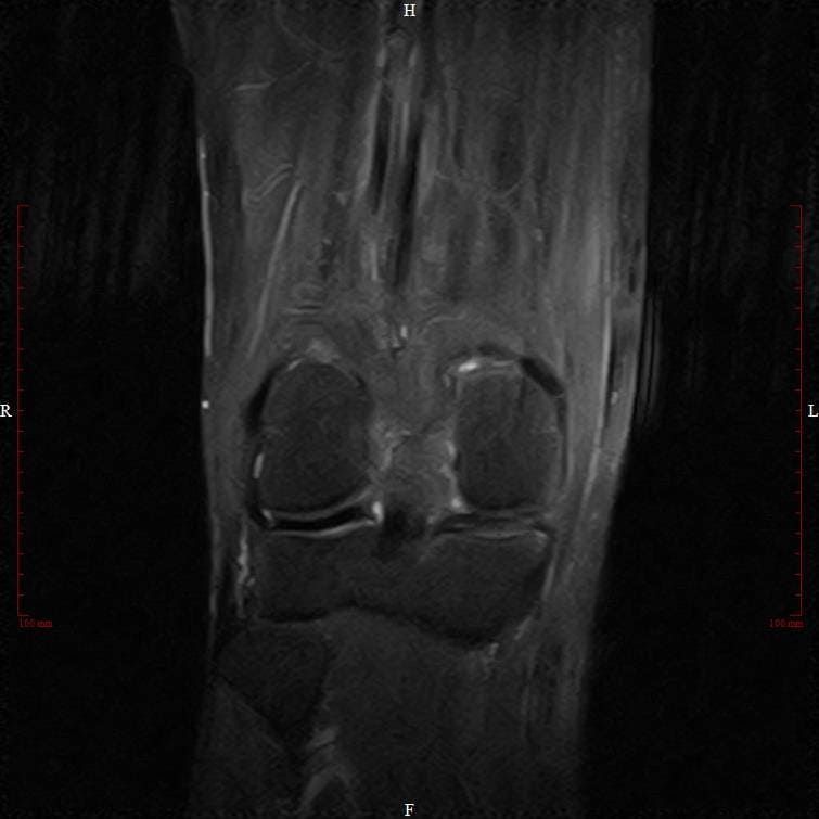
6 months after refixation
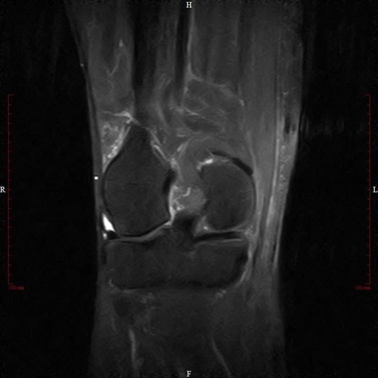
1 year after refixation
These MRI images show almost complete signal normalization in the posterior horn of the medial meniscus after one year. The patient is completely symptom-free.
Our method was nominated for the GERMAN MEDICAL AWARD 2019
View the GERMAN MEDICAL AWARD here
Online appointment
- Schedule now
Im Zollhafen 6, 50678 Köln, Deutschland
