


Magnetic resonance imaging
Magnetic resonance imaging (MRI) is a safe examination method that does not use X-rays and is therefore completely harmless. We use a so-called "open" scanner, meaning patients do not have to go inside a tube. In most cases, contrast medium is also unnecessary.
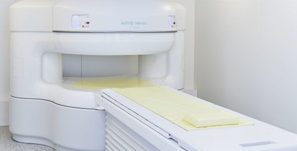
Advantages of an MRI compared to a conventional X-ray
Compared to X-rays, MRI can visualize not only the bony skeleton but also soft tissue and other structures that are not readily visible on X-ray images. This allows injuries or degenerative changes, such as meniscus tears, to be detected at an early stage. Special imaging techniques can also reveal inflammation in MRI scans, often preventing unnecessary surgeries.
In our open device, we also perform injections under MRI guidance. The needle is visible on the MRI image and can therefore be navigated very precisely. This allows joints such as the hip or shoulder to be punctured safely and without exposure to X-rays.
In cases of herniated discs, so-called PDIs (peridural injections) or PRTs (periradicular injections) are performed under MRI guidance near the intervertebral discs.
The following images show some examples from magnetic resonance imaging (MRI), but these only represent excerpts of the examinations. MRI also allows for an almost three-dimensional representation of the human body by combining the individual cross-sectional images.
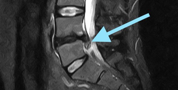
This MRI image of the lumbar spine shows a pronounced herniated disc (blue arrow).
Online appointment
- Schedule now
Im Zollhafen 6, 50678 Köln, Deutschland
