


Osteoarthritis therapy
Osteoarthritis (also known as joint wear or arthrosis deformans) is a painful, progressive joint disease. It results from the permanent wear and tear of the articular cartilage. The hip joint (coxarthrosis), the knee joint (gonarthrosis), and the spine (spondylarthrosis) are most commonly affected. It occurs less frequently in the finger, shoulder, or ankle joints.
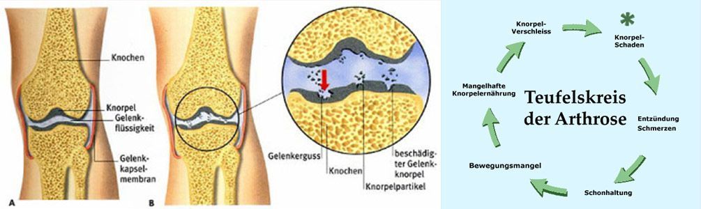
Osteoarthritis can develop at any age, but it occurs much more frequently in older people than in younger people.
The cause is usually seen as an imbalance between the joint's capacity to withstand stress and the actual stress it is subjected to. This can be due to overloading (e.g., increased body weight), congenital or traumatic causes such as joint misalignment, or bony deformities resulting from bone diseases such as osteoporosis.
Overuse leads to cartilage loss at pressure points and a simultaneous decrease in synovial fluid, which restricts the gliding ability of the joint surfaces and irritates the synovium (joint lining) due to constant friction. Furthermore, osteoarthritis can also be a consequence of another condition, such as arthritis (joint inflammation). This is referred to as secondary osteoarthritis. It can also be accompanied by effusion due to overuse, which is then a secondary inflammatory reaction and is called activated osteoarthritis.
A distinction is made between symptoms and complaints in dormant and active osteoarthritis.
In cases of dormant osteoarthritis:
- Feeling of tension and stiffness in the joint
- Often weather-related pain
- Initial pain (at the start of movement), later constant pain during exertion, finally chronic pain
- rubbing noise
- Joint swelling and deformation
- Limited mobility
In cases of activated osteoarthritis:
- Acute inflammation
- Severe joint pain (even at rest)
- Swelling of the joint
- Redness of the skin and increased warmth of the joint
- Protective posture
Osteoarthritis is generally classified as either primary or secondary. Primary osteoarthritis is thought to result from a biological deficiency of the cartilage tissue of unclear origin. Secondary osteoarthritis, on the other hand, arises from mechanical overload, inflammatory changes, or metabolic disorders.
Four different stages are distinguished, which describe the overall damage to the joint:
- Stage I - Early stage: significant cartilage damage (roughness and thinning of the cartilage layer), narrowing of the joint space (visible in X-ray and MRI), changes in the bone beneath the damaged cartilage = often silent osteoarthritis, where patients do not yet experience any symptoms.
- Stage II - Advanced stage: hyaline cartilage is replaced by granulation tissue and inferior fibrocartilage. Pseudocysts form from necrotic cartilage and bone tissue (subchondral cyst), inflammation occurs in the joint area (activated osteoarthritis), and patients experience pain upon weight-bearing and start-up.
- Stage III - Late stage: the cartilage layers are largely destroyed, ulcerations occur, with connective tissue and chondrocytes proliferating (growing), significant inflammation of the joint.
- Stage IV: The bony plate of a joint flattens. Bony outgrowths (osteophytes) form on the bone to absorb the pressure on the joint. Muscle shortening and hardening occur, leading to significant limitations in movement and ultimately to joint stiffness.
There are a number of factors that can contribute to osteoarthritis.
The following potential risk factors are known:
- Overweight
- Sports with a high risk of injury and high impact loads (e.g. football)
- Repetitive adverse joint stresses, e.g. in the workplace (assembly line work, jackhammer, etc.)
- Heavy physical work (mining, shipyards, agriculture, construction), which overloads the joint.
- Hereditary predisposition (familial)
- Lack of exercise leads to poor cartilage nutrition, weak muscle development and consequently, insufficient joint stability.
- Muscular imbalance = imbalance of the muscle groups around the joint
- Advancing age
- Stress caused by pre-arthritic conditions (pre-existing damage that, based on experience, leads to premature joint wear), such as that resulting from a sports injury with cartilage damage to the knee, can also lead to cartilage damage. Incorrect loading, for example due to pronounced bowlegs and the resulting one-sided overload, also leads to cartilage damage.
- Excessive use of the joint, e.g., in a knee joint that has to absorb increased body weight.
- Previous surgeries (e.g., meniscus removal in the knee)
Diagnostic
Various diagnostic procedures are available for diagnosing osteoarthritis. A thorough medical history is a crucial first step in gathering important information about risk factors and the patient's previous disease progression. Imaging techniques such as magnetic resonance imaging (MRI) are also used. The joint space is of particular interest, as it provides insights into the condition of the articular cartilage. Furthermore, MRI can directly visualize cartilage damage.
We are happy to perform MRI scans at any time in our practice – however, please note that we do not offer X-rays.
therapy
There are numerous treatment options that, while they cannot reverse or completely eliminate osteoarthritis, can help slow its progression and alleviate symptoms.
The choice of appropriate treatment depends on the individual form of osteoarthritis, its cause, the stage, and the patient's personal symptoms. The goal of therapy is to reduce pain and limitations in movement and to prevent potential long-term damage.
The following therapies are available in our practice:
- Movement therapy: physiotherapy, Power Plate, Cybex training, biofeedback training
- Various electrotherapies: iontophoresis, galvanization, high-voltage therapy, magnetic field therapy and cryotherapy (cold therapy)
- Hyaluronic acid injections (e.g. Hyalart)
- Autologous EOT II therapy
- Laser therapy
- Kinesio taping
Online appointment
- Schedule now
Im Zollhafen 6, 50678 Köln, Deutschland
